<< Back

2019 Alzheimer's Association Research Fellowship (AARF)

Tau Antibody-Mediated Improvement in Neuronal Function in Tauopathies

Could an advanced brain scan technique reveal how tau antibodies may impact nerve cell function?
 

Qian Wu, Ph.D.
New York University School of Medicine
New York, NY - United States


Background

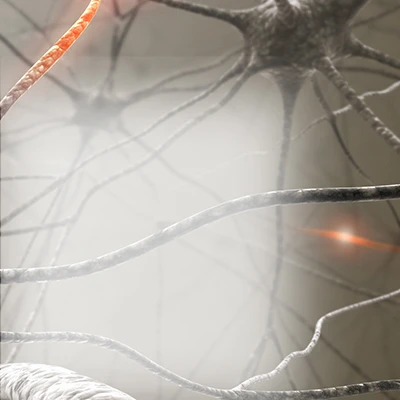
The brain cell’s energy and nutrient transport system is organized in parallel strands like railroad tracks. These tracks allow nutrients to travel across the cell, delivering key materials to the cells, providing them with energy and keeping them healthy. The tau protein helps keep these tracks straight. However, in Alzheimer’s and other brain diseases like frontotemporal dementia and Pick’s disease, the shape of tau protein becomes modified and this could contribute to tau tangles (a hallmark of these diseases) and subsequent nerve cell damage. Although studies have established that different types of tau and its modifications could be a critical in contributing to brain diseases, the biological mechanism by which this occurs is not yet clear.
 
Calcium activity is a key component of brain communication networks and an indicator of nerve cell health. Researchers can measure calcium activity inside nerve cells to understand how well cells are functioning. Past studies have found that abnormal calcium activity may be associated with Alzheimer’s. However the impact of abnormal calcium activity on other brain diseases caused by abnormal tau is not yet clear.
 

Research Plan

Dr. Qian Wu and colleagues will apply a state-of-the-art brain scan technique, called in vivo two-photon calcium imaging, to measure calcium activity inside living mice that have been exposed to tau antibodies. The antibodies are designed to attach to modified tau and then together they would be cleared by the immune system.
 
For the new technique, Dr. Wu will inject genetically-engineered Alzheimer’s-like mice with a calcium “indicator” molecule. The indicator does not change the ability of mice to participate in normal activities and can be seen under specialized brain scans. This will help Dr. Wu’s team to directly observe nerve cell calcium activity in living and awake mice, as opposed to observing them in nerve cells growing in laboratory dishes, or by measuring calcium levels in brain tissue of mice.
 
Dr. Wu’s team will collect calcium activity data in each mouse before, and after, exposing them to the tau antibodies. After collecting the live calcium activity data, Dr. Wu will also collect brain tissue samples from the mice to further analyze calcium activity.
 
Dr. Wu’s overall goal is to understand how the tau antibodies impact nerve cell activity and function in mice that have abnormal tau in their brain.
 

Impact

The results of this study could provide insights into how tau antibodies currently being studied in clinical trials that target abnormal tau may specifically impact nerve cell communication and their function. A better understanding of how these antibodies work could provide valuable insight into their use as potential therapies for brain diseases caused by abnormal tau.
 

Back to Top